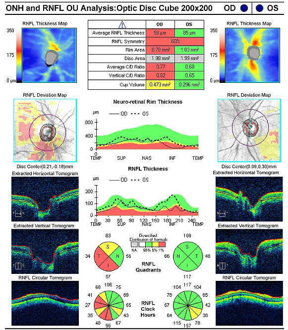


(e) cases in which a more precise diagnosis was not available for any other reason.(d) cases referred elsewhere for investigation or treatment before the diagnosis was made.(c) provisional diagnosis in a patient who failed to return for further investigation or care.(b) signs or symptoms existing at the time of initial encounter that proved to be transient and whose causes could not be determined Retinopathy of prematurity, which can happen in premature babies, causes abnormal blood vessel growth in the retina.(a) cases for which no more specific diagnosis can be made even after all the facts bearing on the case have been investigated.The conditions and signs or symptoms included in categories R00- R94 consist of: In addition, the iWellness retinal scan uses optical tomography (similar to CT scan).8, are generally provided for other relevant symptoms that cannot be allocated elsewhere in the classification. type 2 seen with normal axial length and clinically abnormal sclera and type 3 with normal axial length and clinically normal sclera. 11 OCT-A may show microvascular alterations that are not detected clinically. The Alphabetical Index should be consulted to determine which symptoms and signs are to be allocated here and which to other chapters. OCT cross-section scans may also show regions of inner retinal thinning suggestive of capillary non-perfusion. Practically all categories in the chapter could be designated 'not otherwise specified', 'unknown etiology' or 'transient'. In general, categories in this chapter include the less well-defined conditions and symptoms that, without the necessary study of the case to establish a final diagnosis, point perhaps equally to two or more diseases or to two or more systems of the body. This allows for imaging speeds of 40,000 A-scans. It uses a long-wavelength (near-infrared), broad-bandwidth light source to illuminate the retina and assess the light reflected from retinal tissue interfaces using a spectrometer and Fourier transformation. Signs and symptoms that point rather definitely to a given diagnosis have been assigned to a category in other chapters of the classification. SD-OCT provides two- and three-dimensional images with nearcellular resolution (This chapter includes symptoms, signs, abnormal results of clinical or other investigative procedures, and ill-defined conditions regarding which no diagnosis classifiable elsewhere is recorded.


 0 kommentar(er)
